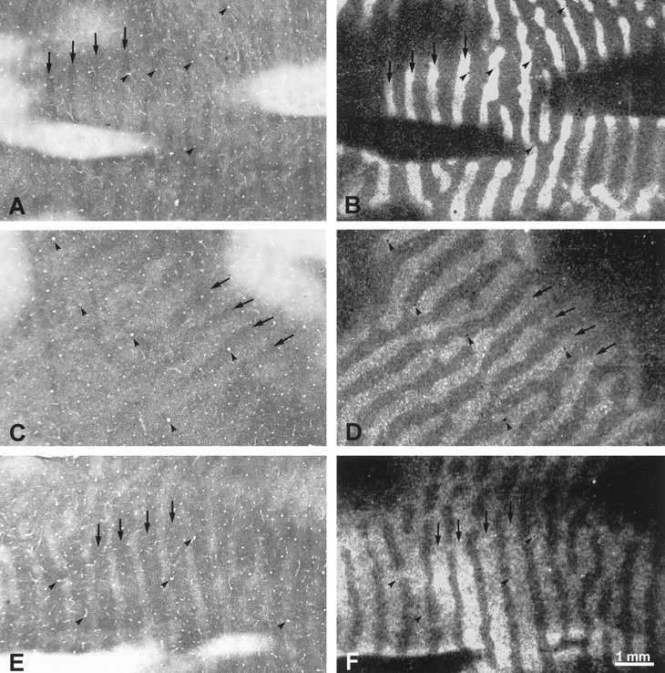
Fig. 5.

Monkey 1 (8° right exotropia). A,B, Adjacent CO and proline sections from the left peripheral cortex (Fig. 3A,B). The autoradiograph is 70 μm deeper than the section illustrated in Figure 3B. The dark CO columns correspond to the left eye’s labeled ocular dominance columns (compare arrows). C,D, Adjacent CO and proline sections from the right operculum (Fig. 3C,D), showing that the dark CO columns belong to the left eye (compare arrows).E, F, Adjacent CO and proline sections, each 70 μm superficial to the sections shown in Figure 3,C and D, again showing a match between the dark CO columns and the left eye’s ocular dominance columns, this time in the right peripheral cortex. In this animal, the dark CO columns were associated with the fixating left eye everywhere in striate cortex of both hemispheres.
