
Fig. 6.
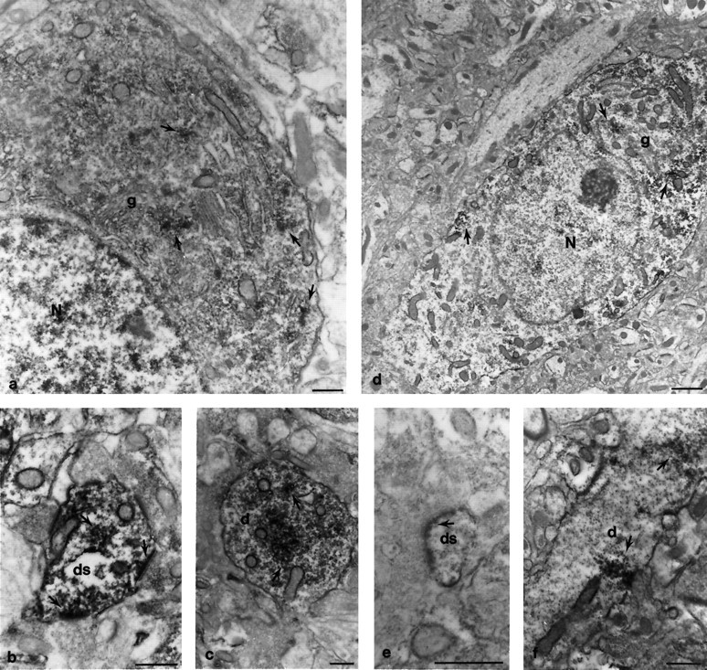
Immunoperoxidase electron microscopic localization of GRIP1 protein in cortex and hippocampus. Coronal rat brain sections were immunostained with the GRIP1-specific antibody and visualized by DAB. The hippocampal CA1 region (a–c) and the cortex (d–f) were analyzed by electron microscopy. The loosely packed, irregular vesicular profiles in the perikaryon and dendritic spines often formed dense patches that were masked with peroxidase product. In the perikaryon of pyramidal cells, the clustered vesicles were often near peri-Golgi complexes, rough endoplasmic reticulum, or scattered in the cytosol (a,d, arrows) and were more condensed in the basal side of neurons (d). Dendritic shafts and large spines contained vesicles with substantial staining (c, f, arrows). Inf, a line of stained vesicles was localized directly underneath the postsynaptic membrane plasma membrane. In large dendritic spines (b, arrow), vesicles were found either clustered in the cytosal or individually merged with stained postsynaptic densities. Some presynaptic nerve terminals were also stained (the terminal in b, right arrow). The electron density of the postsynaptic membrane was significantly enhanced because of immunoperoxidase precipitation. This phenomenon was found in either large dendritic spines (b, arrow) or spine heads (e, arrow). N, Nucleus;g, Golgi complex; d, dendritic shaft;ds, dendritic spine. Scale bar, 0.5 μm.