
Fig. 7.
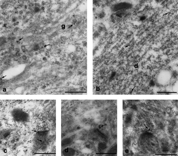
Immunogold electron microscopy localization of GRIP1 protein in CNS. Consistent with results in Figure 6, immunogold particles were found in small clusters, which were frequently associated with vesicular structures and localized near the peri-Golgi and endoplasmic reticulum in the perikaryon (a) and dendritic shafts (b). In b, gold particles outlined a vesicle near microtubule filaments. Gold particles were occasionally found in the PSD (c) but more frequently localized near the PSD (d,e, arrows). Gold labeling was also observed infrequently in terminals (c, bottom arrow). g, Golgi apparatus; d, dendritic shaft. Scale bar, 0.5 μm.