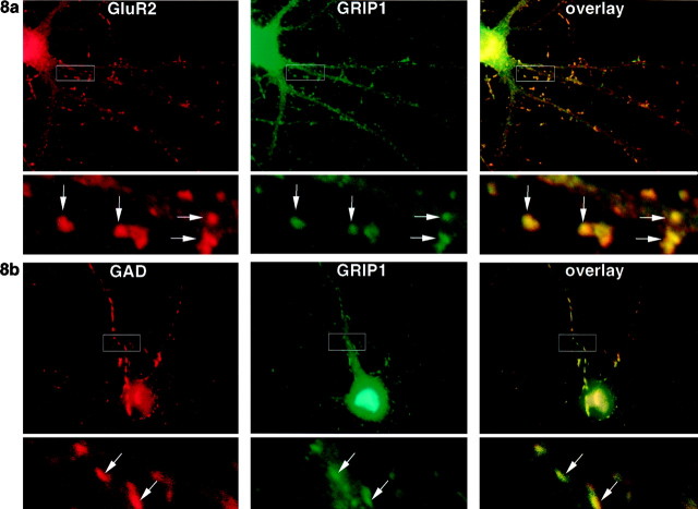
Fig. 8.
GRIP1 colocalizes with AMPA receptors at excitatory synapses and with GAD at inhibitory synapses. Hippocampal neurons in culture (3 weeks) were double-labeled with anti-GRIP1 (green) and monoclonal anti-GluR2 antibodies (a, red), or anti-GRIP1 and monoclonal anti-GAD antibodies (b, red). GRIP1 staining (green) was punctate and was found throughout the neuronal cell body and neurites and colocalized with AMPA at dendritic spines (a, arrows). GRIP1 also colocalized with GAD at inhibitory synapses on dendritic shafts (b, arrow). The bottom panels show an enlarged portion of the area within thewhite rectangles in the top panels.

