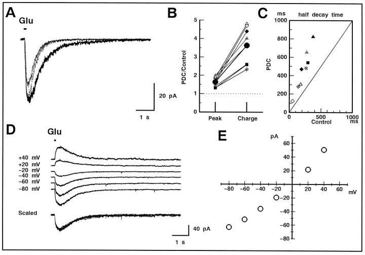
Fig. 3.
Effect of PDC on the non-NMDA receptor-mediated current in GLCs of the retinal slice. A, A GLC was voltage-clamped at −80 mV, and glutamate (200 μm) was puff applied for 100 msec. The tip of the puff pipette was carefully positioned above the IPL to evoke a large response. In both the bath and puff solutions, divalent cations were replaced with Co2+ to suppress the synaptic transmission, andd-AP-5 (50 μm) was included to block the NMDA receptor-mediated current. The glutamate-induced current in the presence of 200 μm PDC (thick trace) was larger in amplitude and decayed more slowly than control (thin trace). After scaling (dotted trace), it is clear that PDC prolonged the glutamate-induced current through non-NMDA receptors. B, Relative increase in the peak amplitude (Peak) and total charge (Charge) before and after the application of PDC. Data were obtained from three cells and are illustrated with different symbols. Duration of the puff was 10 (open), 50 (half-tone), or 100 (filled) msec. The averaged values are shown with large filled circles. C, The scatter diagram illustrates the relationship between the half-decay time of the glutamate-induced current in the absence (Control) and presence of PDC. Allsymbols correspond to those shown in B. D, The current was evoked in a GLC by a 50 msec puff of glutamate in the presence of 200 μm PDC. The holding potential was changed to various values. The current traces are shifted arbitrarily for a better view. All of the current traces were superimposable after scaling (bottom traces).E, The peak amplitude of the glutamate-induced current was plotted against the command potential of GLC. The linear relationship confirmed that the glutamate-induced current was caused by the activation of non-NMDA receptors.

