Fig. 9.

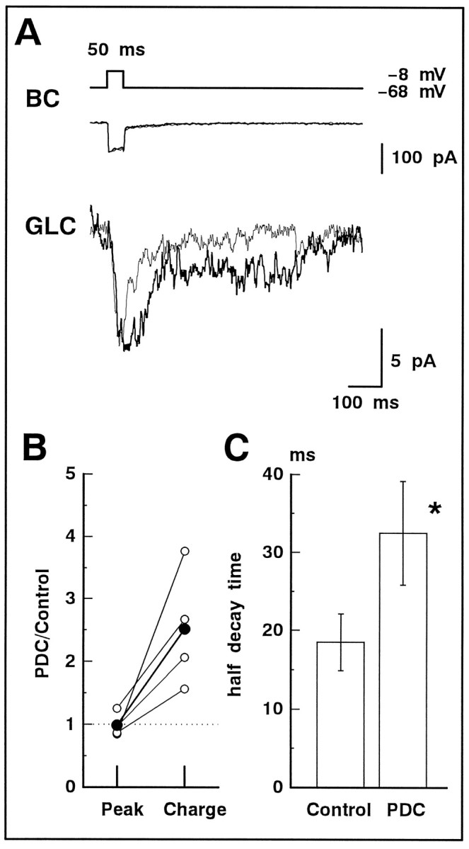
The evoked NMDA-EPSC was prolonged by PDC. A, In the slice preparation, dual whole-cell recordings were performed. A 50 msec depolarizing pulse (from −68 to −8 mV; top) applied to BC activatedICa (middle; thin trace) in BC and evoked the NMDA-EPSC in GLC voltage-clamped at −40 mV (bottom; thin trace). Bath application of 200 μm PDC did not change ICa (middle;thick trace) but prolonged the evoked NMDA-EPSC (bottom; thick trace). The superfusate always contained 5 μmNBQX. B, Relative increase in the peak amplitude (Peak) and total charge (Charge) of the evoked NMDA-EPSC before (Control) and after application of PDC (n = 4). The averaged values are shown by large filled circles. C, The half-decay time of the evoked NMDA-EPSC in the absence (Control) and presence of PDC.
