
Fig. 7.
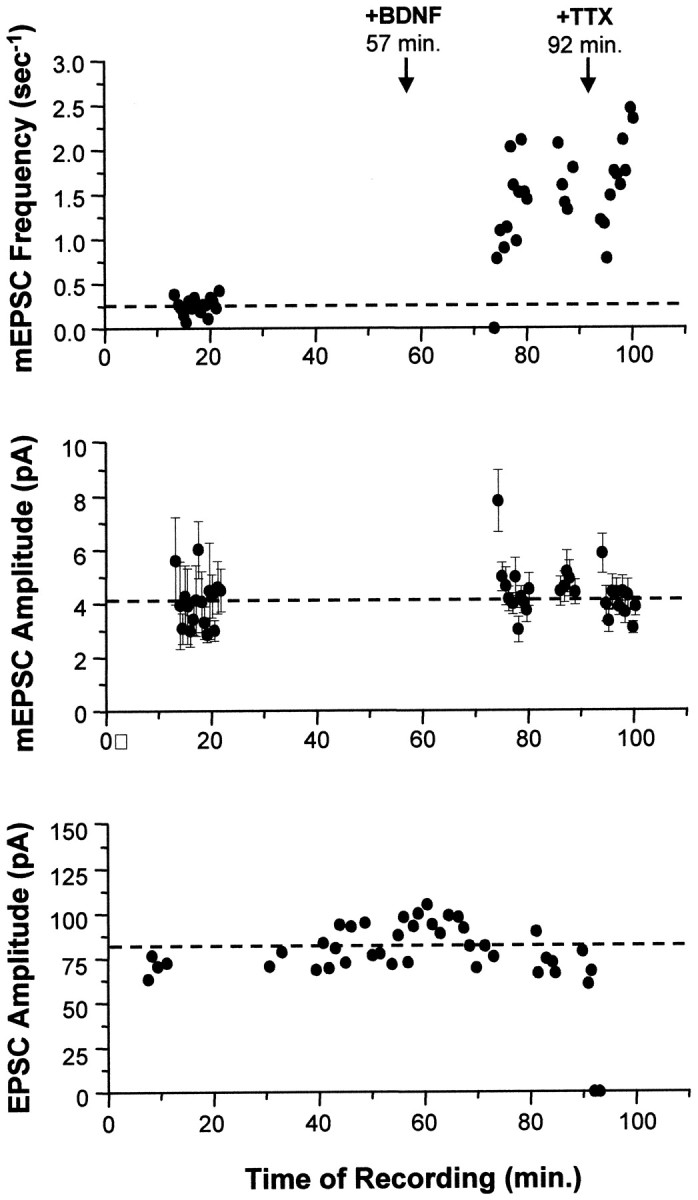
Acute application of BDNF increased only the frequency of mEPSCs. In this example, synaptic currents from a single autaptic neuron were first recorded in the absence of BDNF to establish baseline synaptic transmission properties (mEPSC frequency, mEPSC amplitude, and EPSC amplitude). Each mEPSC point represents 25.6 sec of recording. BDNF was then added to the bath (100 ng/ml att = 57 min), and these synaptic properties were monitored for 35 min. Finally, TTX was added at t = 92 min to confirm that the mEPSCs measured were true action potential-independent minis. Top, The frequency of mEPSCs increased 5.2-fold (p < 2 × 10⁻⁹) in response to BDNF addition. This effect was seen within minutes and was not affected by the subsequent addition of TTX (p > 0.1, comparing average post-BDNF frequencies measured before and after TTX).Center, No differences were observed in average mEPSC amplitudes (±SE) throughout the recording; p > 0.9 for mEPSC amplitudes measured before and after BDNF addition. Blank periods in this and the graph above are due to EPSCs being recorded during these intervals. Bottom, Similarly, EPSCs were unaffected by the addition of BDNF (p > 0.8, comparing pre-BDNF and post-BDNF measurements). Note that the last two EPSC amplitudes are zero, confirming that sodium channels were blocked by TTX, eliminating all action potentials and EPSCs.