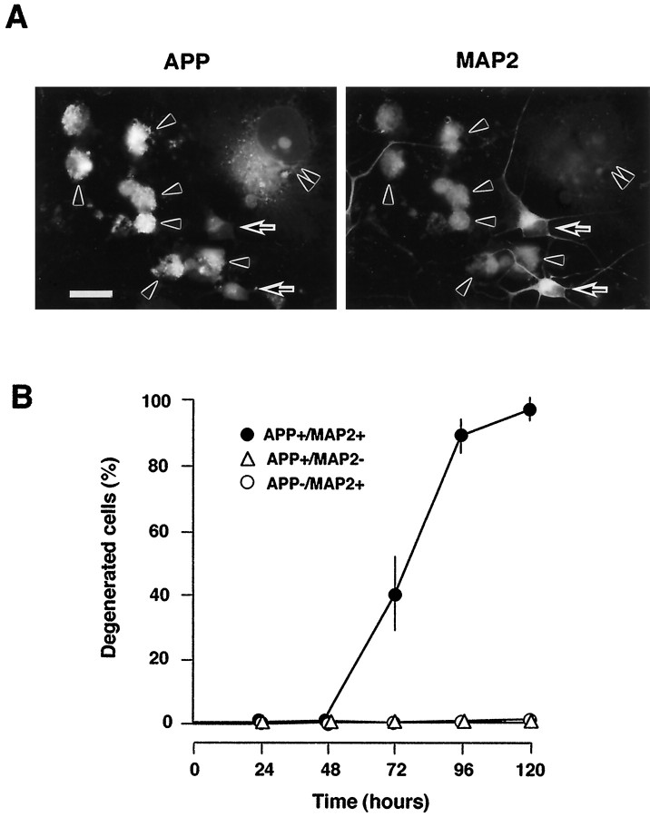
Fig. 2.
Degeneration of AxCAYAP-infected neurons. A, Double immunostaining for APP (left) and MAP2 (right) in AxCAYAP-infected cells at 96 hr. Neurally differentiated NT2 cells were infected with AxCAYAP and fixed 96 hr later. Fixed cells were double stained with an antibody against APP-C terminus (AC1) and anti-MAP2 antibody and visualized by fluorescence microscopy.Arrowheads, APP-accumulating neurons (APP+/MAP2+);double arrowheads, APP-accumulating non-neuronal cells (APP+/MAP2−); arrows, neurons without APP accumulation (APP−/MAP2+). Scale bar, 50 μm. B, Quantification of neurodegeneration. Morphological changes of AxCAYAP-infected NT2 cells were examined at each time point by double immunostaining for APP and MAP2 as shown in A. Degenerated cells showing severe membrane blebbing and complete neurite retraction among 100 cells of each group were counted (mean ± SEM; n = 3).

