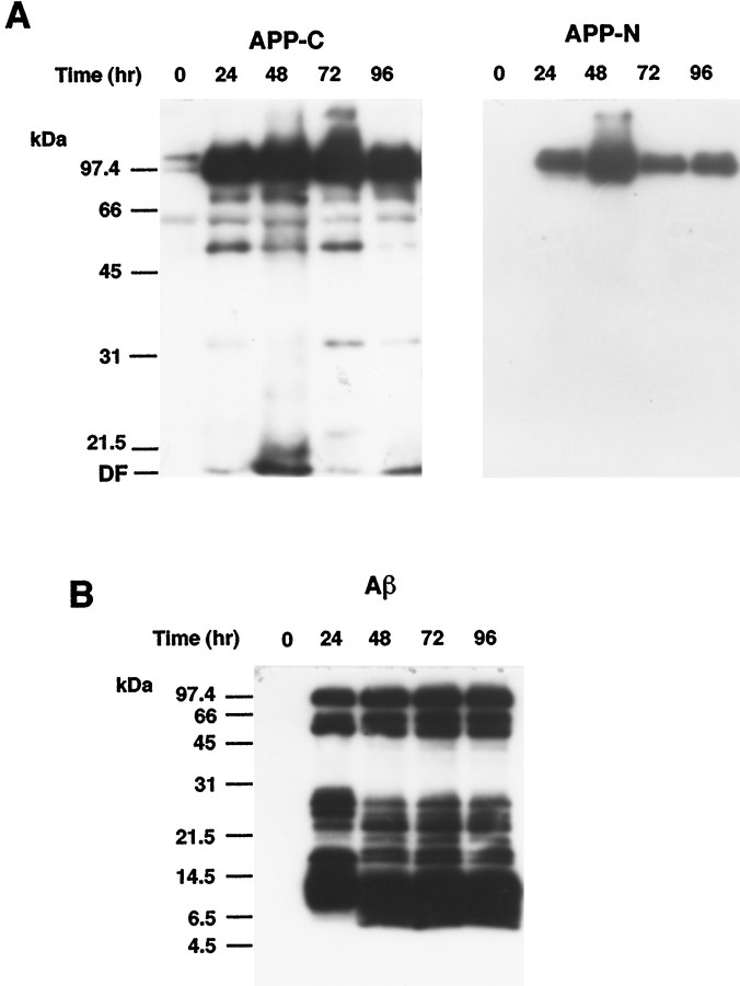
Fig. 3.
Western blot analysis of APP and Aβ peptides in AxCAYAP-infected neurons. A, APP-immunoreactive proteins. Lysates were prepared from enriched neurons at indicated time points after AxCAYAP infection. Proteins (5 μg/lane) were separated by 10% SDS-PAGE and transferred to PVDF membrane. APP C terminus (APP-C, left) and N terminus (APP-N, right) were detected with antibodies AC1 and P2–1, respectively. B, Aβ-immunoreactive peptides (Aβ). Proteins in AxCAYAP-infected neurons (10 μg/lane) were separated by 16% Tris-Tricine SDS-PAGE and transferred to PVDF membrane. After the membrane was boiled in PBS for 5 min, Aβ-immunoreactive peptides were detected with an antibody against Aβ (amino acids 17–24) (4G8). Size markers (in kilodaltons) are on the left.DF, Dye front.

