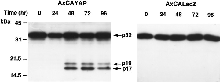
Fig. 5.
Western blot analysis of caspase-3 protein. Enriched neurons infected with AxCAYAP (left) or AxCALacZ (right) were harvested and lysed at each time point indicated. Proteins (20 μg/lane) were separated by 12% SDS-PAGE and transferred to PVDF membrane. Caspase-3-immunoreactive bands were detected with an anti-caspase-3 antibody recognizing both pro-caspase-3 and active subunits. Arrows, Pro-caspase-3 (p32) and its active caspase-3 subunits (p19, p17). Size markers (in kilodaltons) are on the left.

