
Fig. 6.
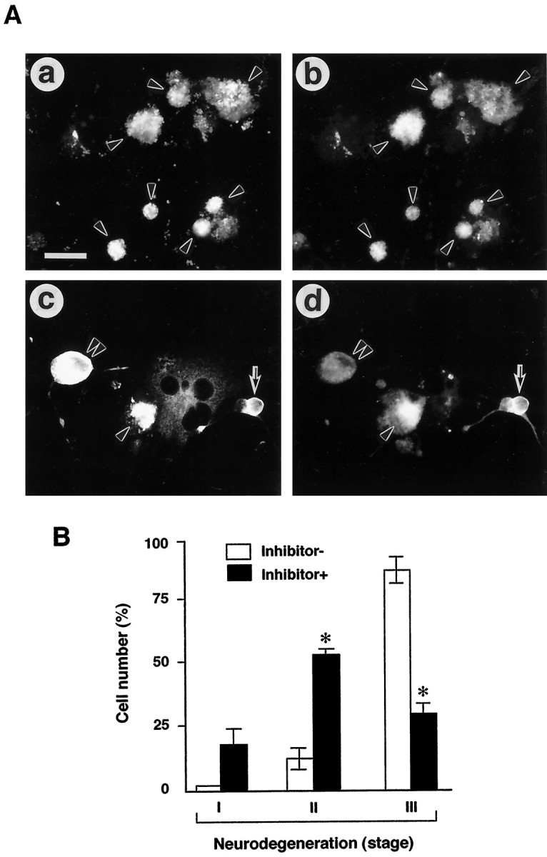
Protection of AxCAYAP-infected neurons from degeneration by caspase-3 inhibitor. AxCAYAP-infected cells were incubated for 96 hr in the absence (Inhibitor−) and presence (Inhibitor+) of 100 μmAc-DEVD-CHO. Cells were fixed and double immunostained for APP C terminus (a, c) and MAP2 (b, d). A, Morphological changes of AxCAYAP-infected neurons in the absence (a,b) and presence (c, d) of the inhibitor. The severity of degeneration displayed by APP-accumulating neurons was classified into three stages: stage I, intact morphology (arrows in c,d); stage II, mild membrane blebbing, disorganized neurites, or swelling of cell body (double arrowheads inc, d); and stage III, severe membrane blebbing and complete neurite retraction (arrowheads ina–d). Scale bar (in a),a–d, 50 μm. B, Quantification of the protective effect of caspase-3 inhibitor on neurodegeneration. AxCAYAP-infected neurons in each group (stages I–III) were counted after double staining for APP and MAP2 (mean ± SEM;n = 3; ≥200 cells per each group). *p < 0.05, significantly different from the Inhibitor− values by Student’s t test.