
Fig. 7.
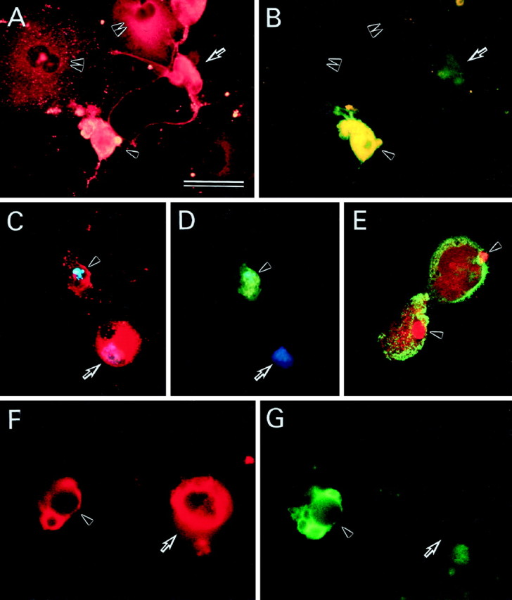
APP and caspase-3 immunoreactivities in AxCAYAP-infected neurons in vitro and in vivo. Neurally differentiated NT2 cells were fixed at 72 hr after AxCAYAP infection and stained for APP (A), activated caspase-3 (B), APP (red) and DNA (blue) (C), activated caspase-3 (green) and DNA (blue) (D), and APP (green) and TUNEL (red) (E). Rat hippocampal tissues were fixed in vivo at 72 hr after AxCAYAP injection and stained for APP (F) and activated caspase-3 (G). AxCAYAP-infected cells were double immunostained for APP N terminus and active caspase-3 subunits with antibodies P2–1 (rhodamine-B) and anti-p20/17 (FITC), respectively (A and B, C andD, and F and G, respectively). The image of chromosomal DNA was superimposed with those of APP and caspase-3 (p20/17) immunoreactivities (C, D). DNA fragmentation and APP were visualized by TUNEL (Texas Red) and immunostaining for APP C terminus with an antibody AC-1 (FITC) by confocal laser scanning fluorescence microscopy (E). See Results for indicated cells. Scale bar (in A), A–D, 50 μm;E–G, 35 μm.