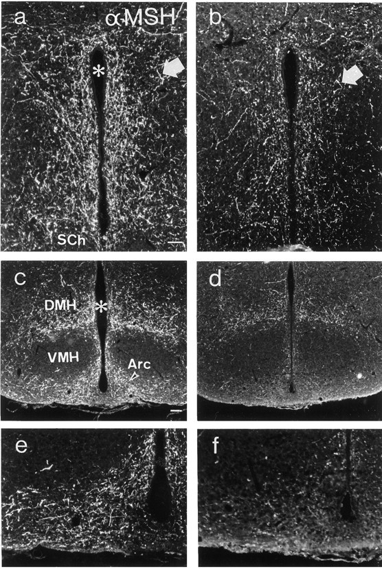
Fig. 5.

Fluorescence micrographs of sections from the periventricular area (a, b) and arcuate nucleus (c–f) of wild-type (a,c, e) andanx/anx (b,d, f) mice stained with antiserum against α-MSH. The number of α-MSH-IR terminals (arrows) are reduced in mutant mice. eand f represent a higher magnification ofc and d, respectively.Asterisk indicates third ventricle. Arc, Arcuate nucleus; DMH, dorsomedial hypothalamic nucleus;SCh, suprachiasmatic nucleus; VMH, ventromedial hypothalamic nucleus. Scale bars: (in a)a, b, e, f, 50 μm; (in c) c, d, 100 μm.
