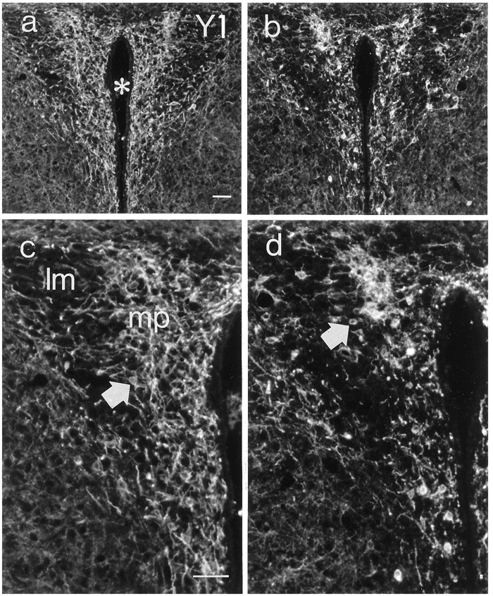
Fig. 8.

Fluorescence micrographs of sections from the paraventricular hypothalamic nucleus of a wild-type (a, c) and ananx/anx (b,d) mouse stained with antiserum against the Y1R. (Micrographs in a and b are magnified inc and d, respectively). There is a reduction in both cell bodies (arrows) and dendritic arborizations. Asterisk indicates third ventricle.mp, Medial parvocellular nucleus; lm, lateral magnocellular nucleus. Scale bar, 50 μm.
