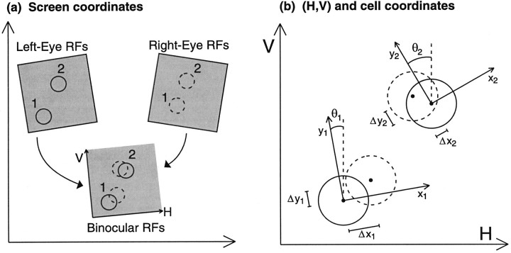
Fig. 2.
a, RFs are initially mapped monocularly through the left and right eyes on the flat screen on which stimuli were projected. Left and right RFs of two cells are shown. Each eye’s RFs, within a small area, may then be mapped onto a common coordinate system, here labeled (H, V), through application of a single, unique transformation operation, consisting of a rotation and translation. b, Binocular RFs of a cell i are most easily described using coordinates (xi,yi) with origin at the center of the left-eye RF and with theyi-axis aligned parallel to the cell’s preferred orientation. The angle θi is defined as the counterclockwise angle from the V axis to the preferred orientation.

