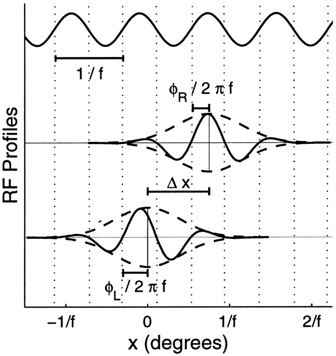
Fig. 4.
Example of a binocular RF. Top, Sinusoid hypothesized by the subregion correspondence model to set absolute phases of both monocular RFs. Middle, bottom, Profiles through the centers of those RFs taken along thex-axis, perpendicular to the cell’s preferred orientation. ON and OFF subregions appear above and below the midlines, respectively, which are displaced vertically by an arbitrary amount for clarity. The Gaussian envelopes (dashed) of the left-eye and right-eye RFs are centered at 0 and Δx. Although the RFs have different phases, φL and φR, measured relative to the centers of their RF envelopes, their zero crossings (dotted lines) occur at identical xvalues; so they can each be described as a Gaussian envelope multiplied by a single sinusoidal function, with wavelength 1/f.

