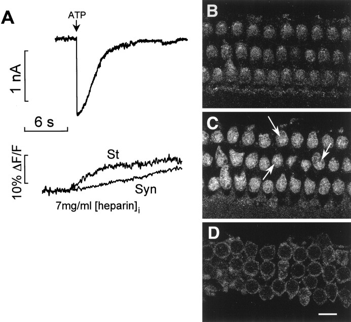
Fig. 5.
Inhibition by heparin and immunolocalization of InsP3 receptors. A, Effect of intracellular heparin (7 mg/ml) on Ca2+ movement.Top, Current evoked by ATP (1 mm, 100 msec);bottom, corresponding fluorescence changes measured in 0 [Ca2+]o from a region adjacent to the OHC cuticular plate (St) and near the synaptic pole (Syn). B—D, Confocal microscopy of ∼0.4 μm cross-sections at 2, 7, and 20 μm below the apical surface of the three rows of OHCs from a whole-mount preparation of the organ of Corti, labeled with the anti-InsP3 receptor antibody. Intense fluorescence was observed just below the cuticular plate (C). Some fluorescence labeling was also detected at the cell cortex along the lateral wall, forming the ring images in D. Scale bar, 10 μm.

