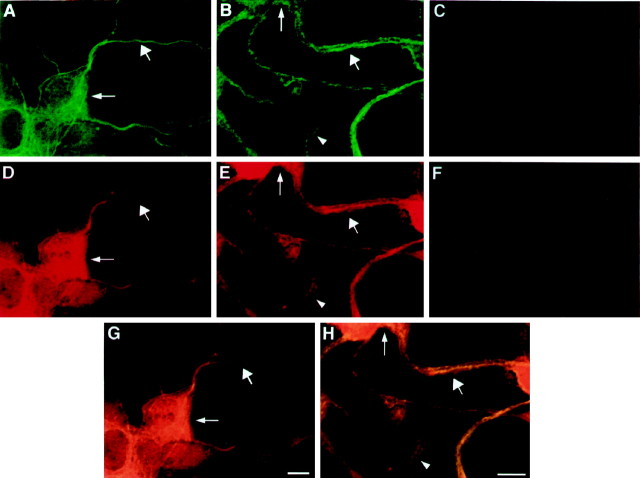
Fig. 5.
Confocal microscopy analysis of PC12 cells stained with tubulin and anti-ELAV-like antibodies. A,B, Confocal image of PC12 cells stained with tubulin antibodies. D, E, Confocal image of PC12 cells stained with anti-Hu serum. C, F, Control experiments showing no penetration of Cy3 or fluorescein signals into the opposite windows. Thin arrow,wide arrow, and arrowhead indicate cell body, neurite, and growth cone, respectively. Magnification: 5× forA, D, G; 6× forB, E, H. Scale bar, 5 μm.

