Fig. 10.
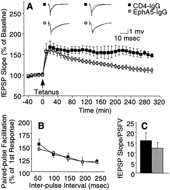
EphA5-IgG impairs LTP maintenance in hippocampal slices prepared from C57BL/6 mice. A, Field EPSP is expressed as percentage of baseline. Twenty sequential responses were averaged and plotted as one point. ANOVA revealed a significant immunoadhesin effect (F(1,8) = 8.09;p = 0.02) and a significant immunoadhesin × time interaction (F(29,232) = 4.98;p < 0.0001). Tukey's HSD test showed that the groups became significantly different (p < 0.05) 90 min after tetanization. Representative traces before and after tetanization at corresponding time points, as indicated, are also shown. B, No immunoadhesin effect was observed on paired pulse facilitation (F(1,11) = 0.164;p > 0.690), which was assessed by applying paired pulses of equivalent intensity at interpulse intervals as indicated. Facilitation ratios are calculated by expressing the slope of the second fEPSP as a percentage of the slope of the first fEPSP.C, Basal synaptic transmission, estimated by ratio of the fEPSP slope to the PSFV amplitude, was not altered by EphA5-IgG-infused (hatched bar) compared with CD4-IgG-infused (black bar) mice (t= 1.119; df = 14; p > 0.28). Estimation of basal synaptic transmission by I/O characteristics using Michaelis–Menten sigmoid curve fit revealed no significant differences (CD4-IgG, mean of 1.30 ± 0.368; EphA5-IgG, mean of 1.54 ± 0.219; p > 0.80).

