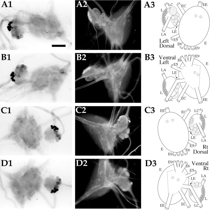
Fig. 8.
AMRP neurons in the pleural and pedal ganglia.A1,In situ hybridization of left ganglion pair dorsal surface. A2,Immunocytochemistry of the left ganglion pair dorsal surface.A3, Drawing of the AMRP neurons on the dorsal surface of the left ganglion pair. B1,In situhybridization of left ganglion pair ventral surface. B2,Immunocytochemistry of the left ganglion pair ventral surface.B3, Drawing of the AMRP neurons on the ventral surface of the left ganglion pair. C1,In situhybridization of right ganglion pair dorsal surface. C2,Immunocytochemistry of the right ganglion pair dorsal surface.C3, Drawing of the AMRP neurons on the dorsal surface of the right ganglion pair. D1,In situhybridization of right ganglion pair ventral surface.D2, Immunocytochemistry of the right ganglion pair ventral surface. D3, Drawing of the AMRP neurons on the ventral surface of the right ganglion pair. L, Pleural ganglion; E, pedal ganglion; LE,pleuropedal connective; EE, pedal commissure;EC, cerebropedal connective; LC,cerebropleural connective; LA, pleuroabdominal connective; E5, posterior tegumentary nerve (P5);E6, anterior parapodial nerve (P6); E9,posterior pedal nerve (P9). Not all nerves are drawn for simplicity. Scale bar: A1, 500 μm (same in all panels).

