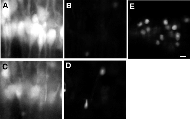
Fig. 6.
MEQ fluorescent cells within area CA1 pyramidal cell layer exclude PI 10 min after H2O2. An MEQ-loaded slice was constantly superfused with PI.A, MEQ fluorescent neurons in area CA1 before H2O2. B, Neurons inA imaged with the rhodamine filter for PI.C, MEQ fluorescent neurons 10 min after H2O2. D, Neurons inC imaged with the rhodamine filter for PI.E, As a positive control, the slice was exposed to methanol for 2 min to allow entry of PI into the same cells shown inA–D. All images were at a 1.7× electronic zoom to maintain registry between multiple laser lines. Scale bar, 20 μm.

