
Fig. 4.
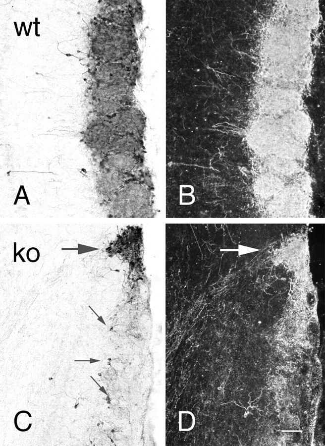
Bright-field (A,C) and dark-field (B,D) photomicrographs of TH immunostaining of olfactory bulb in wild-type (A,B; wt) and OCNC1-null (C,D; ko) mice. In wild-type mice, strong staining is observed in periglomerular cells and glomerular processes of all glomeruli. In the null mice, only scattered periglomerular cells (small arrows) contain TH in most glomeruli and, as illustrated in dark-field (D), labeled processes are absent. In contrast, TH staining is normal in both cells and processes in the atypical or necklace glomeruli (large arrow). Scale bar, 40 μm.