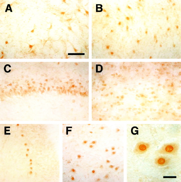
Fig. 7.
Immunohistochemistry of KKIAMRE labeled with an anti-KKIAMRE antibody. KKIAMRE expression was detected in neurons of various brain regions: interpositus nucleus (A) and dentate nucleus (B) in the cerebellar deep nuclei, hippocampal CA3 (C), cerebral cortex (D), cerebellar Purkinje cell (E), and facial nuclei in the brain stem (F). G shows immunoreactive neurons in the facial nuclei with a higher magnification having a strong signal in the nucleus. Scale bars: A–F, 100 μm; G, 25 μm.

