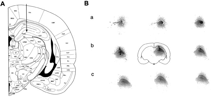
Fig. 1.
A, Cannula/bipolar electrode assembly location is indicated by arrow.B, Nine entire coronal sections labeled by33P GluR2(B) AS-ODN autoradiography are on display (as indicated by outline of center section) from the most anterior portion of the dorsal hippocampus to the ventral posterior region. Distance between sections on display is 600–800 μm (a–c) so that the infused GluR2(B) AS-ODN spread or regional accumulation was approximately 2.4–2.8 mm3within the ipsilateral dorsal but not ventral hippocampus. Mediolateral gradients diminished in posterior sections (B,c), and ODNs did not spread to the contralateral hippocampus or to other brain regions (not visible or seen aswhite background).

