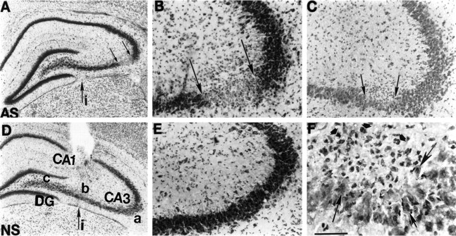
Fig. 3.
Photomicrographs depicting GluR2(B) knockdown-induced cell loss and morphological alterations in CA3 hippocampal neurons after 4 d of infusions. A, Thionin-stained section from GluR2(B) AS-ODN-treated pup at low magnification shows marked loss of CA3a pyramidal neurons (betweensmall arrows) just after the bend and posterior to the level of infusion site (large arrow). B, Loss of CA3a neurons by thionin is shown at higher magnification.C, F, Hematoxylin/eosin-stained section 360 μm posterior to section displayed in B at low (C) and high (F) magnifications reveal eosinophilic neurons (large arrows) and apparent glial cell proliferation (short arrows) in posterior sections around the distant lesion.D, NS-ODN control section at level of infusion site and at low and high (E) magnifications shows that CA3 neurons distant from cannula infusion site are intact with normal cytoarchitecture. Scale bar, 50 μm. i, Infusion site;AS, antisense; NS, nonsense.

