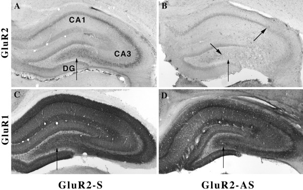
Fig. 7.
A, Photomicrograph showing that GluR2(B) immunolabeling (6C4) of CA1–CA3 stratum pyramidale was dense and uniform; GluR2(B) antibodies predominantly label soma, whereas GluR1(A) antisera label soma and dendrites. A, In sense-treated animal sections, GluR2(B) immunolabel was dense and evenly distributed throughout the cytoplasm of CA3 neurons.B, After GluR2(B) hippocampal knockdown (bold arrowhead), GluR2(B) immunoreactivity was markedly reduced throughout the CA2, CA3a–b, and part of CA3c subregions (betweenarrows); CA1 and DG were downregulated but to a lesser extent. C, GluR1(A) immunoreactivity after S-ODNs was intense throughout the hippocampus. D, GluR1(A) immunoreactivity was unchanged, suggesting that an increase in the GluR1(A)/GluR2(B) protein was achieved by the GluR2(B) knockdown.

