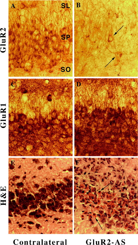
Fig. 8.

Photomicrographs of GluR2(B) and GluR1(A) immunolabeling of CA3 neurons after GluR2(B) knockdown at high magnification (400×). A, In control GluR2(B) sections, robust labeling of soma and proximal dendrites was observed. B, CA3 neurons were particularly decreased in GluR2(B) immunoreactivity; a few neurons were immunopositive near the lesion (arrows). C, GluR1(A) control immunoreactivity was dense and continuous with apical and basilar dendrites. D, GluR1(A) immunoreactivity near the lesion was unchanged. E, Control hematoxylin/eosin stain of CA3 just before the bend from contralateral hippocampus. F, Shrunken nuclei and eosinophilia of CA3a neurons were detected by hematoxylin/eosin stain (arrows). SR, Stratum radiatum; SP, stratum pyramidale;SO, stratum oriens.
