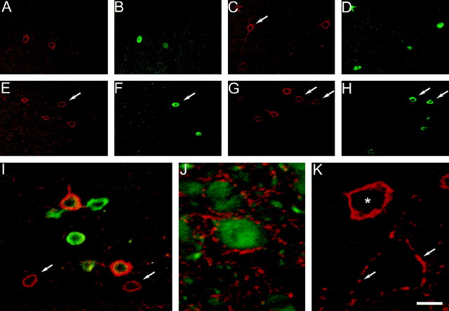
Fig. 7.
Coexpression of Kv3.2 and PV, calbindin, or somatostatin. A–H, Confocal immunofluorescence images of superficial (A, B) and deep (C–H) cortical layers in somatosensory cortex double labeled for Kv3.2 and PV (A–D), Kv3.2 and calbindin (E, F), and Kv3.2 and somatostatin (G, H).Red fluorescence, Kv3.2; greenfluorescence, PV, calbindin, or somatostatin. A,B, The two Kv3.2-labeled cells in superficial cortical layers shown in A are PV-positive (B). C, D, There are four Kv3.2-labeled somata in this image obtained in deep layer V. Three of these show PV immunoreactivity (D), whereas one (C, arrow) is not stained for PV. E, F, Four Kv3.2-labeled neurons in layer VI, one of which (arrows) shows calbindin immunoreactivity. G, H, Two of six Kv3.2-labeled cells in this field are somatostatin-positive (arrows). I, Confocal immunofluorescence image of deep cortical layers in somatosensory cortex triple labeled for Kv3.2, PV, and calbindin. The immunofluorescence for Kv3.2 (red) and PV or calbindin (green) has been superimposed. Two Kv3.2-stained cells in this image have no green fluorescence (arrows). J, Confocal immunofluorescence image of somatosensory cortex (layer VI) double labeled for calbindin (green) and Kv3.2 (red). Note Kv3.2-immunoreactive puncta surrounding calbindin-positive neurons. K, Confocal immunofluorescence image of layer V somatosensory cortex labeled with antibodies to Kv3.2. Shown is an unstained pyramidal neuron surrounded by Kv3.2-positive puncta (some of which are indicated byarrows) near a Kv3.2-labeled cell (asterisk). Scale bar: A–H, 30 μm;I, 12 μm; J, K, 7.5 μm.

