
Fig. 5.
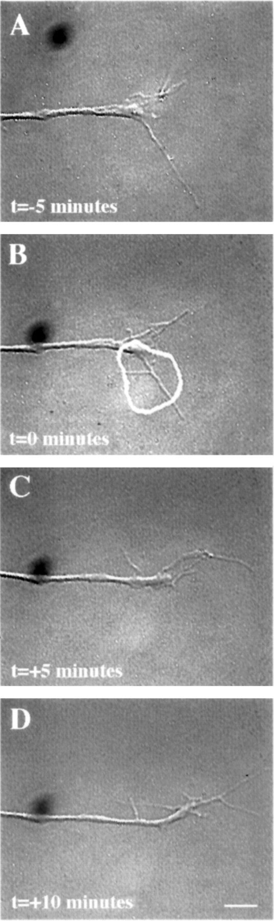
Micro-CALI of the intracellular domain of NCAM 180 using MG-labeled 4d caused regional growth cone retraction.A, A growth cone was first observed for 5 min. A region of the growth cone was chosen for micro-CALI (white outline), laser irradiation was initiated att = 0 (B) and continued untilt = +5 min (C), and observed for 5 more minutes (D). In C, the filopodia and lamellipodia of the irradiated region retracted from the laser spot, whereas the rest of the growth cone did not appear to be affected. In D, the growth cone began to grow in another direction, causing a visible bend in the neurite. The time-lapse imaging was done using DIC microscopy instead of phase contrast as was used for Figures 4 and 6. Scale bar, 10 μm.