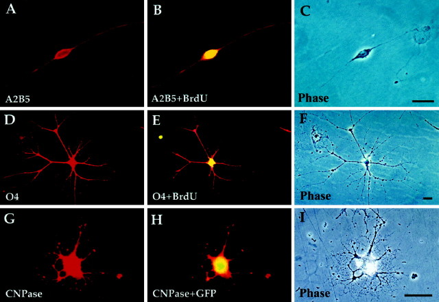
Fig. 6.
P/hCNP2:hGFP-sorted cells divide and express oligodendrocytic markers. A–C, A bipolar A2B5+/BrdU+ cell 48 hr after FACS. D–F, Within 3 weeks, the bipolar cells matured into fibrous, O4+ cells. These cells often incorporated BrdU, indicating their in vitro origin from replicating A2B5+ cells. G–I, A multipolar oligodendrocyte expressing CNP, still expressing GFP 3 weeks after FACS. Scale bar, 20 μm.

