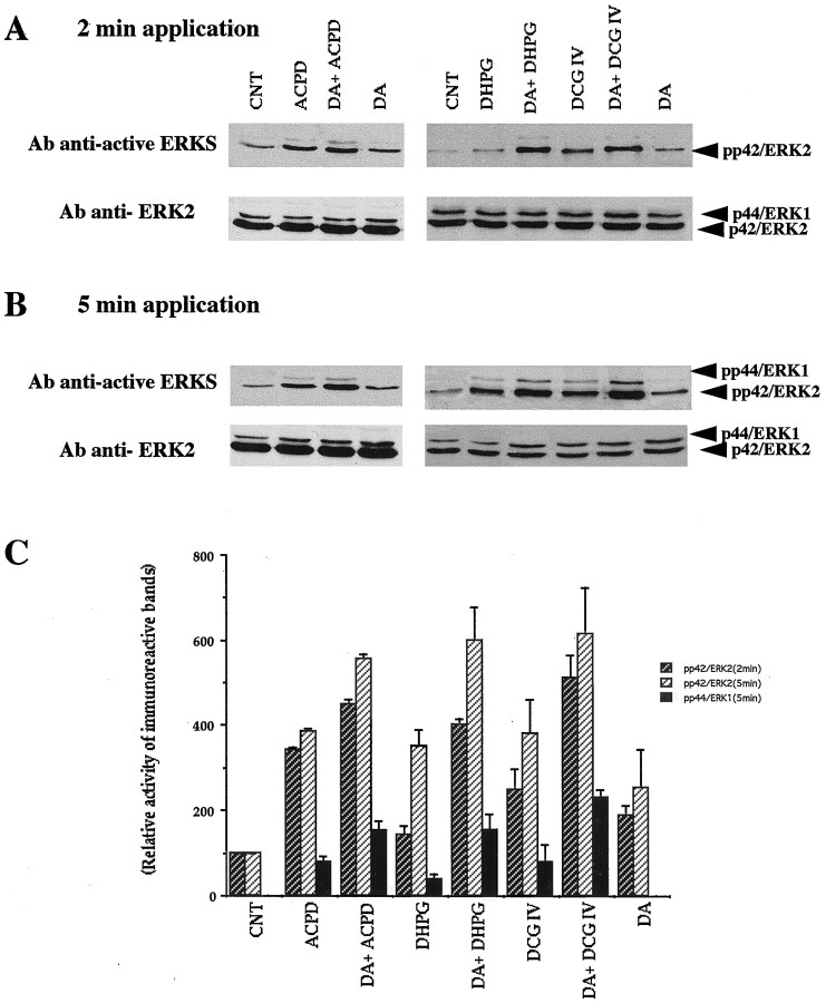
Fig. 11.
Dopamine and mGluR agonists additively or synergistically activate MAP-Ks as detected with anti-active MAP-K antibody. Slices were incubated at 28°C in buffer containing 1 μm bicuculline without other drugs (CNT) or with dopamine (DA, 100 μm) or an mGluR agonist (1S,3R-ACPD, 100 μm; DHPG, 100 μm; DCG IV, 100 nm) or both for 2 or 5 min. Bicuculline itself did not change MAP-K activity (data not shown). Equal amounts of lysates (50 μg) were resolved on a 10% acrylamide gel. Activated MAP-Ks were detected with an antibody anti-active MAP-Ks (anti-active ERKs) that recognizes dually phosphorylated ERKs. After stripping, the immunoblot was reprobed with polyclonal anti-ERK2 (see Materials and Methods). A,B, Results with 2 and 5 min application, respectively. Note that phosphorylated protein bands are always denser after dopamine plus an mGluR agonist than either drug alone (see Results).C, Quantification by scanning densitometry of the autoradiogram of anti-active ERKs using NIH Image 1.6. The data are representative of at least three independent experiments. For the ERK2 activity (2 and 5 min), every drug condition showed statistical significance of at least p < 0.05 over control. For the ERK1 activity (5 min), the increase seen in the presence of dopamine and a given mGluR agonist was always significantly larger than the increase seen in the presence of that mGluR agonist alone (p < 0.05). The graph is plotted as percentage changes relative to CNT levels for p42/ERK2, and raw arbitrary numbers for p44/ERK1 because of the lack of p44/ERK1 phosphorylation in CNT.

