
Fig. 1.
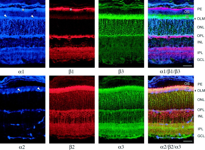
Confocal fluorescent micrographs of adult mouse retina sections triple-labeled with α1, β1, and β3 (top row) or α2, β2, and α3(bottom row) isoform-specific primary antibodies and Cy5-conjugated (blue, anti-mouse), TRITC-conjugated (red, anti-rat), or FITC-conjugated (green, anti-rabbit) secondary antibodies. The images on the right side include all three color channels. Arrowheads and asterisksindicate Müller cell endfeet in the region of theOLM and label in the retinal pigment epithelium, respectively. Scale bar, 50 μm.