Fig. 1.
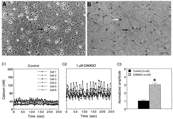
Effect of DAMGO on baseline intracellular Ca2+ oscillations in cultured hippocampal neurons. Shown are phase contrast (A) and bright-field (B) micrographs of hippocampal neurons in cultures immunostained with an antibody to the neurotransmitter GABA. The cultures contain a variety of neuronal types based on morphological characteristics and immunostaining for GABA, glutamate, and peptides (e.g., somatostatin; D. Gruol, unpublished data). The neurons grow on a background of astrocytes, identified by immunostaining with an antibody to GFAP. The white arrow points to a neuron immunostained with an antibody for GABA; the dark arrowpoints to a neuron that did not immunostain with the antibody. Calibration bar, 60 μm. C1, C2, Representative recordings of intracellular Ca2+ oscillations in six cultured hippocampal neurons in a microscopic field (different field than in A) under baseline control conditions (C1) and after the addition of DAMGO to the bath saline (C2). Each neuron is represented by a different symbol. Small intracellular Ca2+ oscillations were observed under baseline conditions, and these oscillations were enhanced dramatically in amplitude by the addition of DAMGO to the bath saline. DAMGO also synchronized the oscillations among the neurons in the field. C3, Mean values ± SEM for the amplitude of the intracellular Ca2+ oscillations (peak to trough measurement) in a population of control and DAMGO-treated neurons in the same culture dish as the recordings shown in C1 andC2. *Significant difference from control (p < 0.05; ANOVA). In these and all other experiments the Mg2+ concentration of the bath saline was 30 μm, and the bath saline contained glycine (5 μm).

