
Fig. 3.
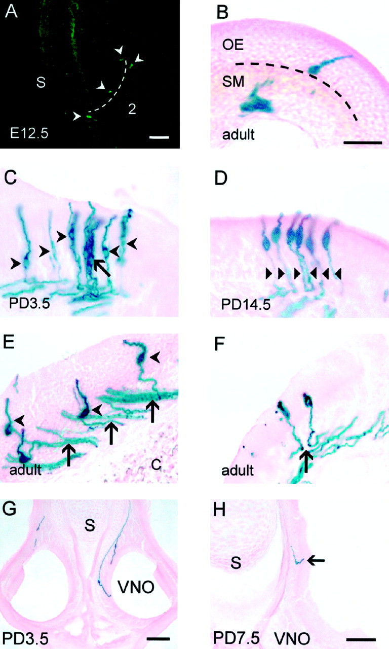
Analysis of the distribution of P2 neurons and their axonal projections. Coronal cryostat sections were stained using X-gal immunohistochemistry (A) and X-gal histochemistry (B–F, I).A, P2 was expressed in neurons (arrowheads) found in zone three (indicated bybroken line) of the olfactory neuroepithelium at E12.5.B, A lone P2 neuron with its cell body at the base of the olfactory neuroepithelium in adult. C, A cluster of P2 neurons at PD3.5 (arrow) are distinct from adjacent individual neurons (arrowheads). Individual neurons were identified by focusing through the thick section. D, Axons expressing P2 do not fasciculate (arrowheads) as they pass through the olfactory neuroepithelium (PD14.5).E, Once P2 axons have exited the olfactory neuroepithelium, they form numerous small bundles within the submucosa (arrows) in adult. Arrowheads depict widely separated perikarya. F, Adjacent P2 neurons send their axons in opposite directions (arrow) in the submucosa (adult). G, H, P2 neurons in other sensory neuroepithelia. Coronal cryostat sections were stained using X-gal histochemistry. G, Two neurons expressing P2 are seen in the vomeronasal organ (PD3.5). H, At PD7.5, a P2 neurons is present in the organ of Masera (arrow). Scale bar, 100 μm. C, Cartilage; OE, olfactory epithelium; S, nasal septum;SM, submucosa; VNO, vomeronasal organ;2, ectoturbinate 2.