
Fig. 4.
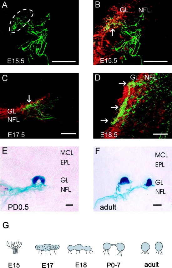
Development of glomeruli in the olfactory bulb. A–D, Double-label immunohistochemistry of coronal cryostat sections stained for β-galactosidase (green) and OMP (red). A single image for β-galactosidase is presented in A, and merged images for OMP and β-galactosidase are presented in B–D. B is the same section as A. E, F, Coronal cryostat sections stained with X-gal histochemistry. A, Confocal image of β-galactosidase at E15.5 reveals P2 axons are diffusely dispersed within a localized area of the olfactory bulb (dotted lines). B, Identical tissue section asA, disclosing both OMP and β-galactosidase, shows dispersed axons in the glomerular layer (arrow).C, By E17.5, P2 axons have begun to condense in the glomerular layer. D, At E18.5, individual glomeruli become visible (arrows) but remain connected by large bundles of axons. E, A glomerulus adopts a rounded morphology by PD0.5, although it remains joined by a band of axons.F, In the adult, glomeruli are completely separated.G, Schematic representation of double-glomerular development. Scale bar, 50 μm. EPL, External plexiform layer; GL, glomerular layer; MCL, mitral cell layer; NFL, nerve fiber layer.