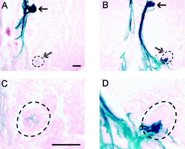
Fig. 5.
A–D, Partial innervation of glomeruli by P2 axons. Coronal cryostat sections stained for X-gal histochemistry. C and D are high-power images of A and B, respectively.A, C, P2 axons are seen to completely innervate the target medial glomerulus (filled arrow), whereas P2 axons are dispersed throughout a subregion of the extra glomerulus (open arrow). B,D, A subregion of the extra glomerulus is innervated by P2 axons (the glomerular border is defined by a broken line). Scale bar, 100 μm.

