
Fig. 6.
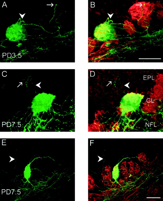
A–F, Behavior of axons targeting glomeruli in the early postnatal period. Coronal cryostat sections immunostained for β-galactosidase (green) and OMP (red). A, C, andE are single images of β-galactosidase staining only;B, D, and F are merged images of β-galactosidase and OMP. A,B, At PD3.5, the growth cone of a P2 axon (arrow) is seen at the surface of an adjacent P2-negative glomerulus. Axons loop back into the target glomerulus (arrowhead). C, D, At PD7.5, a P2 axon exits the target glomerulus (arrowhead), enters the mitral cell layer, and turns back to the glomerular layer. This axon also branches deep to the glomerular layer (arrow). E,F, At PD7.5, a P2 axon (arrowhead) passes through the glomerular layer and heads toward the mitral cell layer. Scale bars, 100 μm. EPL, External plexiform layer;GL, glomerular layer; NFL, nerve fiber layer.