
Fig. 5.
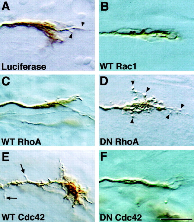
Rho-family GTPase mutants influence growth cone morphology. Shown are RGC growth cones in sections ofXenopus brain immunostained with anti-luciferase (A) or anti-myc antibodies (B–F). A, A growth cone transfected with the control protein luciferase;arrowheads indicate filopodia. B–F, Growth cones transfected with wt or dn mutant GTPases.Arrowheads in D indicate the abnormal, balled filopodia observed on dn-RhoA-expressing growth cones.Arrows in E indicate backbranches observed on axons of wt-Cdc42-expressing cells. Notice also the unusually large growth cone. Scale bar, 15 μm.