
Fig. 2.
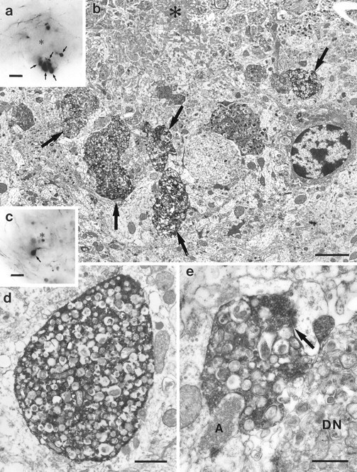
Ultrastructure of PHAL-labeled dystrophic entorhinal boutons in the hippocampus. a, Light micrograph of an amyloid plaque (asterisk) in the molecular layer of the dentate gyrus surrounded by numerous large PHAL-labeled entorhinal boutons (arrows).b, Electron micrograph of the bottom half of the plaque shown in a. The amyloid (asterisk) is surrounded by several dystrophic PHAL-labeled entorhinal boutons (arrows). c, Light micrograph of an amyloid plaque (asterisk) in the molecular layer of the dentate gyrus. The arrow points to a heavily PHAL-labeled entorhinal bouton. d, Electron micrograph of the PHAL-labeled entorhinal bouton illustrated in c. The bouton is filled with numerous multilamellar bodies, characteristic of dystrophic neurites. These structures are surrounded by electron dense immunoprecipitate. e, PHAL-labeled entorhinal bouton in the vicinity of a plaque. This dystrophic bouton contains synaptic vesicles and forms a synapse with a spine. Thearrow points to the synaptic cleft. An unlabeled dystrophic neurite (DN), as well as a normal axon terminal (A) are also illustrated. Scale bars:a, c, 10 μm; b, 2.5 μm; d, 1 μm; e, 0.5 μm.