
Fig. 5.
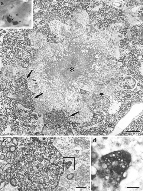
Ultrastructure of entorhinal dystrophic boutons in the white matter. a, Light micrograph of an amyloid plaque (asterisk) in the alveus of the hippocampus. Three PHAL-labeled entorhinal boutons surround the plaque (arrows). b, Electron micrograph of the plaque illustrated in a. The amyloid core (asterisk) is surrounded by numerous dystrophic neurites. The three large entorhinal boutons illustrated ina are indicated by arrows. One of the unlabeled dystrophic neurites is myelinated (short arrow). c, Serial section of the middle PHAL-labeled bouton illustrated in b. The heavily immunolabeled bouton is filled with multilamellar bodies.d, Serial section of the rectangleillustrated in c. This part of the dystrophic bouton shows elements of synaptic specialization, i.e., clustered vesicles. Scale bars: a, 10 μm; b, 5 μm;c, 1 μm; d, 0.25 μm.