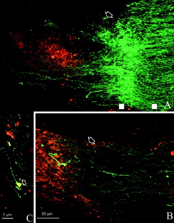
Fig. 5.

High magnification of bcl-2 (A) and wt (B) nerves illustrated at the site of crush. Arrows indicate the lesion site. Note the wealth of sprouting profiles in the bcl-2 (A) compared with the paucity of fibers present in the wild type (B). White squares are examples of the points intersected by lines perpendicular to the nerve major axes and used to calculate the density values of fluorescence staining. The square on theleft is at the sprouting site, and theright one is located more proximally. C, The tip of a regenerating fiber from A exhibits the typical morphology of a growth cone (arrow).
