
Fig. 1.
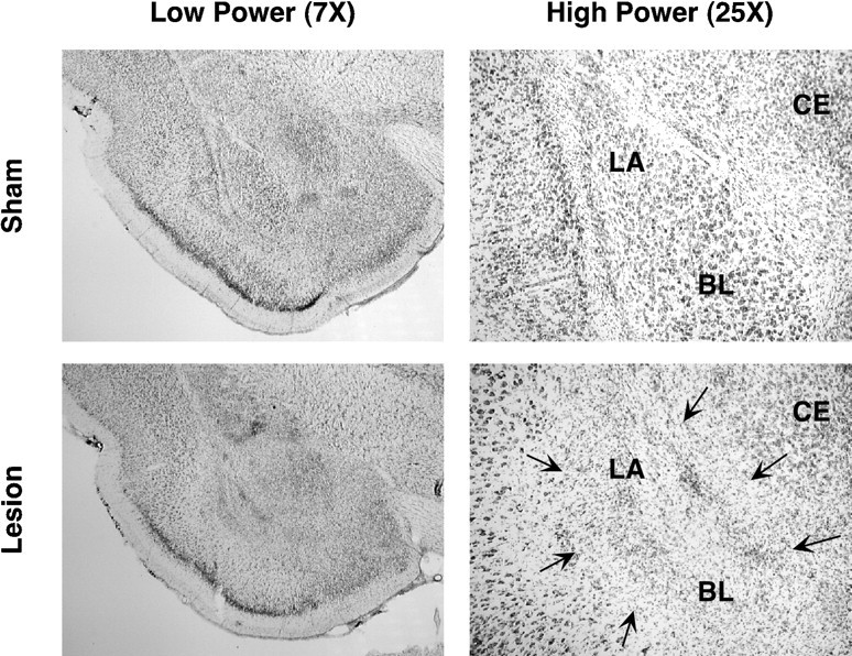
Photomicrographs of thionin-stained brain sections at low and high magnifications from representative rats receiving either sham surgery or a neurotoxic BLA lesion. The lesion (indicated by arrows) was confined to the basolateral amygdaloid complex. BL, basolateral; CE, central nuclei of the amygdala; LA, lateral nuclei of the amygdala.