
Fig. 1.
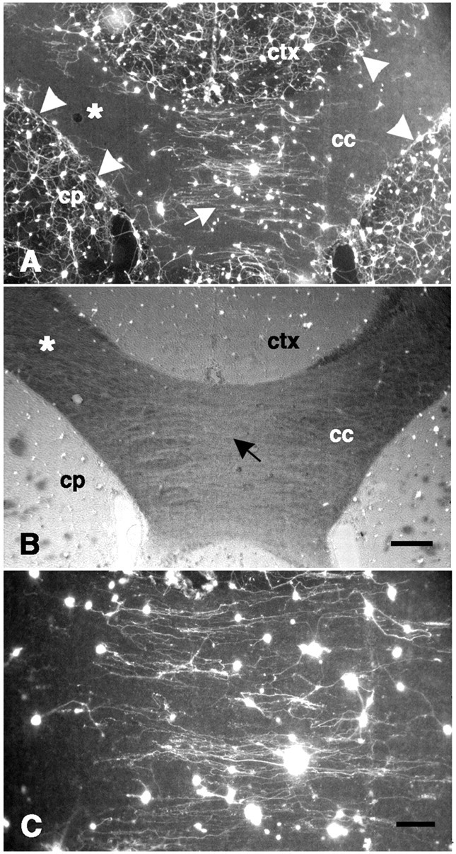
Dissociated neurons on a coronal forebrain section. A, The density of fluorescence-labeled neurons is greatest on gray matter, such as the neocortex (ctx) and caudate-putamen (cp); intermediate levels of attachment are found near the corpus callosum (cc) midline (white arrow); and virtually no neurons are attached to more lateral portions of the corpus callosum (white asterisk). The dense plexus of neurites on gray matter is sharply inhibited at the border with the corpus callosum (white arrowheads). From neurons attached to the corpus callosum, neurite outgrowth is oriented in a direction parallel to the longitudinal axis of the tract (see C) in contrast to the complex pattern of neurite outgrowth on gray matter (see also Fig. 2A). B, The same field shown in A is shown with fluorescence in combination with phase-contrast optics. Note the low optical density (black arrow) near the midline of the corpus callosum corresponding to the relatively parallel orientation of the fibers here, in contrast to the darker regions (white asterisk) found laterally where fibers run more obliquely.C, Higher power photomicrograph of the center of fieldA showing neurite outgrowth that is primarily in parallel with the underlying fiber tract is shown. Scale bars:A, B, 300 μm; C, 100 μm.