
Fig. 4.
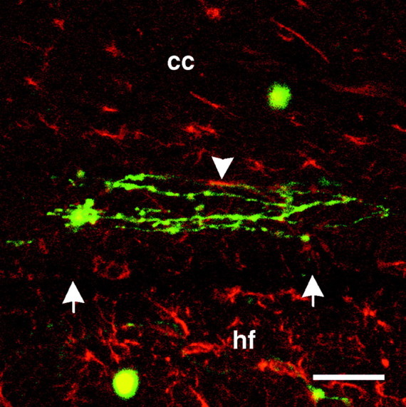
Composite confocal photomicrograph of dissociated neurons on GFAP-labeled sections. Dissociated neurons and neurites (green) on corpus callosum (cc) near the border (white arrows) with the underlying hippocampal formation (hf) are shown. GFAP immunoreactivity is shown in red. Colocalization of neurites and GFAP immunoreactivity is limited (white arrowhead). Scale bar, 50 μm.