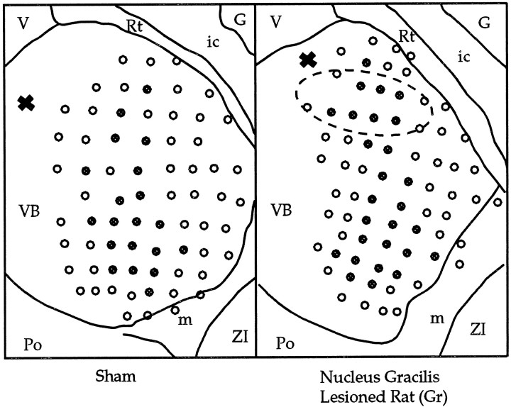
Fig. 7.
Reconstructions of thalamic electrode penetrations for a Sham and Gr animal are presented, viewed dorsally. Tissue was sectioned in the horizontal plane and Nissl-stained. An electrolytic lesion (X) at the top left of each schematic served as a reference point along the dorsoventral axis. The focal zone of plasticity of the VPL is indicated (ellipse). All electrode tracks that encountered sites responsive to shoulder stimulation are indicated by filled circles. G, Globus pallidus; ic, internal capsule; m, medial lemniscus;Po, posterior thalamic nuclear group; Rt, reticular thalamic nucleus; V, third ventricle;ZI, zona incerta.

