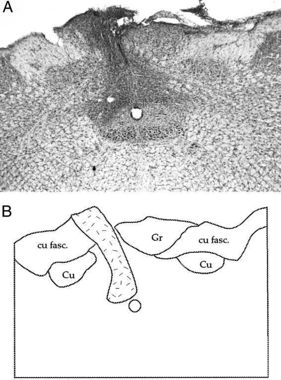
Fig. 8.

A, Photomicrograph of a nucleus gracilis lesion; B, an accompanying schematic: nucleus gracilis (Gr), nucleus cuneatus (Cu), and cuneatus fasciculus (cu fasc.). A dense gliotic zone demarcates what remains of the lesioned gracile nucleus (stippled area). Such damage was observed along the length of nucleus gracilis.
