
Fig. 2.
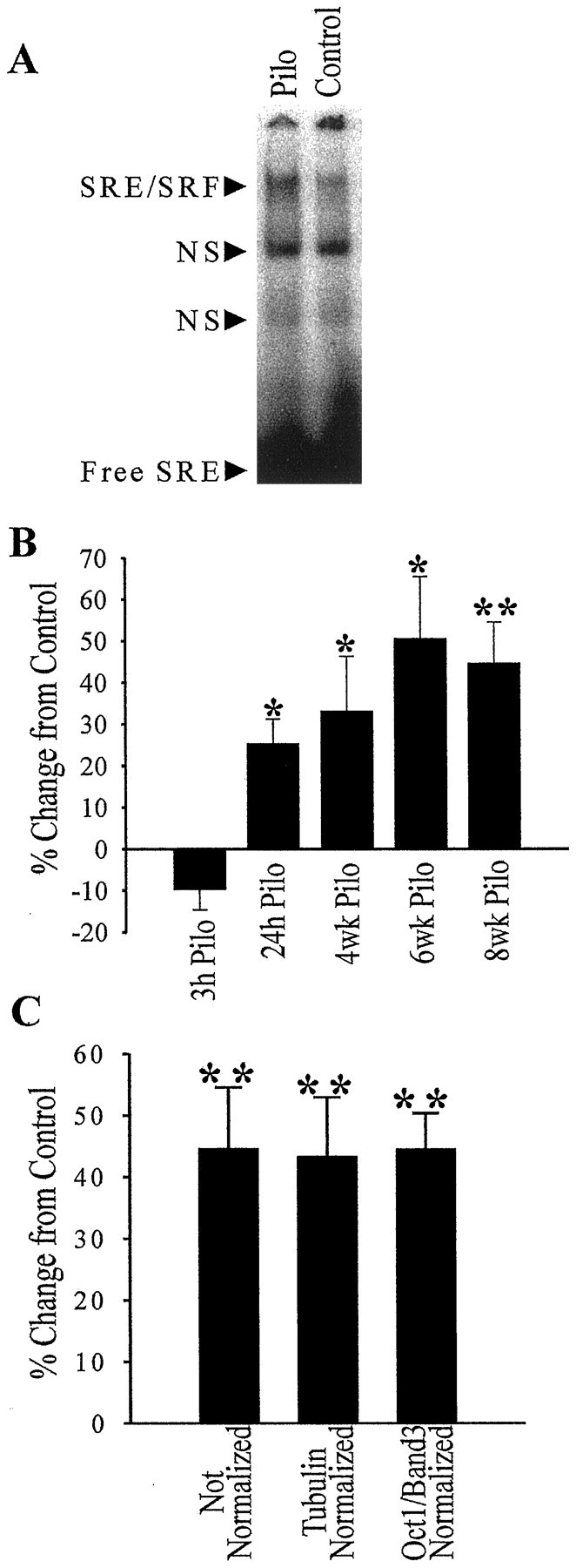
Specific binding of SRF protein to its SRE DNA element is induced during epileptogenesis. EMSAs were conducted in the same manner as in the control reactions in Figure 1, except that the gels were visualized and quantitated by phosphor imaging. A representative EMSA using 8 week post-treatment hippocampal nuclear-enriched fractions from pilocarpine-treated (Pilo) and saline-treated (Control) rats is shown (A). Quantitation of these gels and determination of percent change in SRE binding over paired control nuclear-enriched fractions for five time points (B) revealed a significant increase in SRF binding acutely at 24 hr (n = 6) and at 4 (n = 4), 6 (n = 4), and 8 (n = 6) weeks. The 8 week pilocarpine EMSA data that was not normalized was compared with the data (C) that was normalized by either tubulin protein levels in these nuclear-enriched fractions or EMSA data for transcription factor binding that did not show any changes (Oct1 band three). Error bars represent SEM; *p < 0.05; **p < 0.01; Student’s ttest.