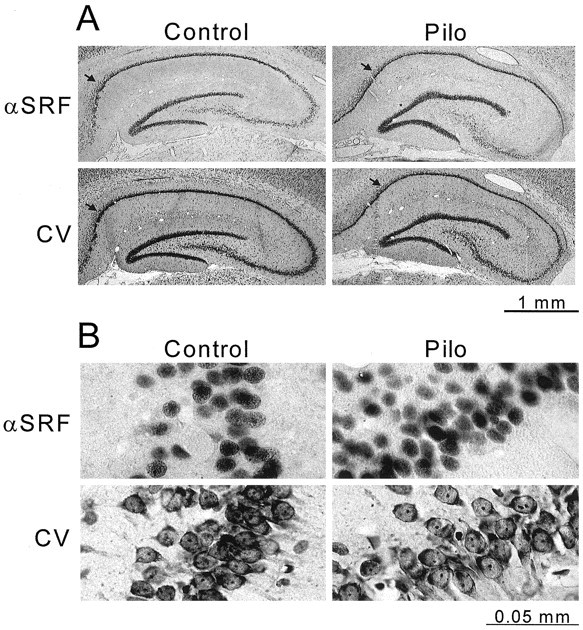
Fig. 7.

SRF immunoreactivity is localized to the nuclei of the neuronal cell regions of the epileptic (Pilo) and control rat hippocampus. Adjacent coronal sections were sliced from a paraformaldehyde-fixed, paraffin-embedded brain of control saline-injected (left panels) and pilocarpine-injected (right panels) rats at 8 weeks after treatment. SRF immunoreactivity (αSRF) was visualized using a DAB substrate kit (see Materials and Methods). Cell bodies in adjacent slices were stained with cresyl violet (CV). Images of cresyl violet- and SRF-stained slides were captured using a digital camera. Low-magnification views of control and epileptic hippocampi are shown in A. Higher magnification views are presented below in B of the CA1 hippocampal region, designated by arrows in A. Scale bars below A and B indicate actual size.
