
Fig. 2.
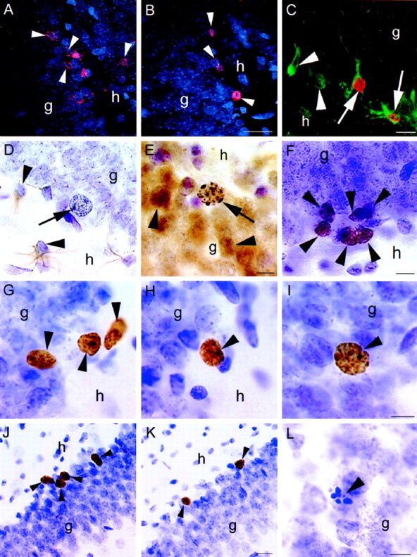
Confocal laser-scanning images of BrdU-labeled cells (arrowheads) in the dentate gyrus of female (A) and male (B) rats injected with BrdU five times and perfused 2 d after the last injection. More BrdU-labeled cells were observed in females compared with males. C, Confocal image of BrdU-labeled cells colabeled with TOAD-64, a marker of immature neurons (arrow), in the dentate gyrus of an adult female.Arrowheads indicate TOAD-64-labeled cells not labeled with BrdU. D, Light microscopic image of a BrdU-labeled cell (arrow) that is not colabeled with GFAP, a marker of astroglia, in the dentate gyrus of an adult female.Arrowheads indicate GFAP-labeled cells not labeled with BrdU. E, BrdU-labeled cell (arrow) that is colabeled with calbindin, a marker of mature neurons, in the dentate gyrus of an adult female. Arrowheads indicate calbindin-labeled cells not labeled with BrdU. F, Ki67-labeled cells (arrowheads) in the subgranular zone of the dentate gyrus. These cells were similar in morphology and location to BrdU-labeled cells at early time points. BrdU-labeled cells (arrowheads) at 4 d (G), 7 d (H), and 21 d (I) after BrdU injection in the dentate gyrus of the adult female. With increasing time after BrdU injection, many BrdU-labeled cells become incorporated into the granule cell layer and express the morphological characteristics of mature granule neurons. More BrdU-labeled cells (arrowheads) were observed in animals injected during proestrus (J) compared with those injected during estrus (K). These animals were perfused 2 hr after injection with BrdU. L, Pyknotic cell (arrowhead) in the dentate gyrus of an adult female. These cells were more numerous in females than males during the majority of the estrous cycle, g, Granule cell layer;h, hilus. Scale bars: B, 25 μm (applies to A, B); C, 10 μm;E, 10 μm (applies to D,E); F, 10 μm; I, 10 μm (applies to G–I); K, 25 μm (applies toJ, K); L, 10 μm.