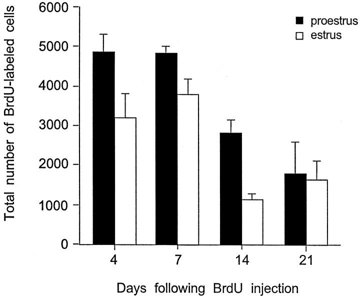
Fig. 4.
Stereological estimates of the total number of BrdU-labeled cells in the dentate gyrus of adult female rats 4, 7, 14, and 21 d after a single BrdU injection administered during proestrus (black bars) or estrus (white bars). The numbers of BrdU-labeled cells decreased over time in both groups but were greater in females injected during proestrus up to 14 d after BrdU labeling. By 21 d, this difference was no longer detectable. Bars represent mean + SEM each obtained from three animals. Asterisk indicates significant difference from proestrus (p < 0.05).

